This is the webpage for the Turku PET Centre neuroimaging course organised in 2021. The course has ended, but all materials will remain freely available. You can view PDF versions of the presentations by clicking on the lecture title, and the recorded lectures are available on Echo360 platform via this link. See below for information on how to access the sample datasets.
Principles And Applications of Neuroimaging Data Analysis
Turku PET Centre neuroscientists are happy to announce that the second edition of our PET-MRI neuroimaging course will be held online August 18.-20. 2021. This course will give a hands-on overview on the basic brain imaging analysis techniques applicable to PET as well as structural and functional MRI data. The course is aimed at PhD students, post-docs, and scientists working on medical, cognitive, and computational neuroimaging. Participants are assumed to have rudimentary knowledge of neuroimaging and working with PET and MRI data, but this is not strictly necessary. After the course, the participants should be able to independently process and analyse the data that has been retrieved from PET camera / MRI scanner. Analytic pipeline will focus on the PET Centre brain data analysis framework but can be adapted to any analysis platform.
The course is arranged outside the formal University of Turku degree system so we cannot give study credits directly for the participation. We will however give certificate (on request) of participation stating that the course covers approximately 2 ECTS credits. Pleaser contact prof. Lauri Nummenmaa for details.
Registering
Registration for the course is closed. You can however view the recorded lectures using the link below.
Practical information
Due to the current COVID regulations at the University of Turku, the course will be organised fully online using Echo360 platform. It is strongly recommended that you use a desktop computer or laptop (rather than tablet computer) for viewing the lectures so you to can follow the online and hands-on demonstrations.
You can access the course site by clicking this link
All lectures (classes) will be streamed live according to the schedule below. Lecture duration is 35 minutes +10 minutes for questions and speaker change. When logging on to Echo360, select the the currently active class to view the live stream. Not that this requires that you actively move from class to the next when the current class ends. Lectures will be stored permanently and they can also be viewed afterwards, the recordings are available almost immediately after the live stream ends.
Asking questions
You can ask questions from the speaker using Discord. Click here to login to our course channel. Unfortunately the Echo360 discussion forum does not work for attendants who do not have an account at our university, so we have to use another service. Because the lectures will be recorded, the speakers will discuss the questions and comments after rather than during each lecture. We will do best to answer all the questions posed in the chat tool – if not immediately, at least after the lecture.
Schedule
Note – schedule is in Helsinki time (EEST), which is UTC+3. Changes are still possible so please re-check the schedule on the webpage and on Echo360. Links below lead to the PDF versions of the slides, use the link above for accessing the recorded lectures.
Wednesday August 18th: Principles of brain imaging
8:45-10:00 Basic principles of human neuroimaging (Lauri Nummenmaa)
10:00-10:45 Preprocessing volumometric brain imaging data (Jouni Tuisku), exercise
10:45-11:30 Pharmakokinetic modelling of PET data: Principles (Jussi Hirvonen)
11:30-13:00 Lunch break
13:00-13:45 Pharmakokinetic modelling of PET data: Applications with MAGIA pipeline (Jouni Tuisku), example code
13:45-14:30 Using Carimas software for image analysis (Chunlei Han)
14:30-15:00 Coffee break
15:00-15:45 Second level analysis of PET data (Lauri Nummenmaa)
Thursday August 19th: Practical issues in brain imaging and analysis
8:15-9:00 Physiology of the BOLD signal and image acquisition (Kerttu Seppälä)
9:00-9:45 GLM for fMRI analysis (Kerttu Seppälä)
9:45-10:00 Coffee break
10:00-10:45 Experimental designs for fMRI (Lauri Nummenmaa)
10:45-11:30 Preprocessing with fMRIprep (Vesa Putkinen)
11:30-13:00 Lunch break
13:00-13:45 First level models (Lihua Sun)
13:45-14:30 Second level models (Severi Santavirta), exercise
14:30-15:00 Coffee break
15:00-15:45 Data visualization (Vesa Putkinen)
Friday August 20th: Advanced topics in brain signal analysis
8:15-9:00 Bayesian statistical inference (Tuulia Malen)
9:00-9:45 Statistical Pattern Recognition with fMRI (Heini Saarimäki)
9:45-10:00 Coffee break
10:00-10:45 Connectivity analysis (Ana Triana Hoyos)
10:45-11:30 Model-free analysis of BOLD-fMRI data (Severi Santavirta)
11:30-13:00 Lunch break
13:00-13:45 Neuroinformatics and large-scale analysis (Lauri Nummenmaa)
13:45-14:30 Wrap-up (All speakers)
Virtual lecture by Mr Malmsteen can be viewed here
Data packages used in the course
PET dataset
This is the full NRM dataset with the LondonPride ligand, kindly provided by the King’s College London PET team. The dataset consists of two PET scans (and associated files) per subject: a baseline scan and a displacement scan. The dataset is available from NITRC via this link.
Pet dataset for MAGIA
This is a subset of the LondonPride dataset for learning MAGIA, the in-house data processing pipeline hosted by Turku PET Centre. Download the dataset from this link.
fMRI dataset
This small dataset from Turku PET Centre contains EPI and T1 data from an experiment, where the subjects viewed pictures with varying content. Download dataset from this link.
LINKS TO SOFTWARE PAGES
MAGIA toolbox
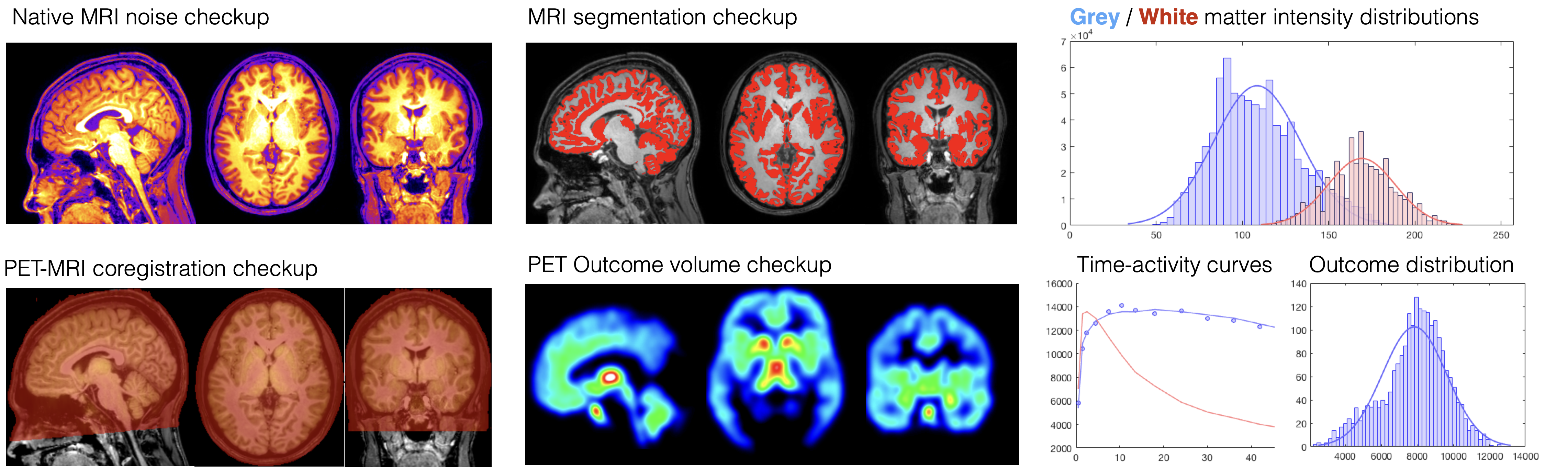
MAGIA is a comprehensive brain-PET modelling suite, which has been optimised for large-scale brain-PET analysis. MAGIA is installed on the Yngwie neuroimaging server and thus available to PET Centre neuroscientists. If you want to install magia on your own computing framework, you can download the source from our git repository.
Data analysis software and tools
SPM12: Multimodal (MRI / PET / EEG / MEG) data processing and analysis suite, runs on Matlab
FSL: Standalone MRI data processing and analysis suite
Freesurfer: Standalone tool for analysis and visualization of structural and functional neuroimaging. Most well known for the excellent cortical surface mesh generation algorithms
AFNI: Standalone MRI data processing and analysis suite
BrainVoyager: Standalone commercial MRI data processing and analysis suite
PMOD: Standalone commerical PET modelling and image processing tool
DTI tools
DTIprep: General-purpose too for DTI analysis
FSL: has also good tensor fitting packages as well as tools for tract-based analysis
ExploreDTI: a graphical toolbox for tractography
Volumometric image viewer tools
Carimas: Medical image analysis platform developed at Turku PET Centre. Capable of visualization, segmentation and modelling of multiple types of medical images.
MRIcron: Image viewer with basic analytic, image manipulation and ROI drawing capabilities, a reliable workhorse
Mango: Advanced volumometric viewer with image editing and rendering capabilities. A nice companion for mricron.
Freeview: Built-in viewer for Fressurfer. Excellent for viewing 2D and 3D images in tandem
FSLeyes: Built-in image viewer for FSL. Good capabilities for volume viewing and support for 4D visualisation.
FSLview: Previous version of FSLeyes.
3D rendering tools
Freeview: Built-in viewer for Freesurfer. Excellent for viewing 2D and 3D images in tandem
Surf Ice: fast and efficient surface rendering tool, highly recommended if you want to work using GUI
3dSlicer: Open-source software package for visualization and medical image computing
Caret: Multi-purpose surface visualization and analysis tool. No longer updated, integrated into connector workbench
MRIcroGL: Graphially more advanced and faster version of MRIcron with better 3D habdling capabilities
FSLview can also do some 3D image rendering