On this page you will find snapshots to the work of the Emotion Lab in engaging and accessible format though videos and images. All items are presented for non-commercial and educational purposes only. The original publications may be downloaded from our Publications page.
Videos
Welcome to the Emotion Lab!
Let’s take a ride towards the fascinating world of emotions and imaging science! Emotion Lab is one of the world’s leading research groups in imaging and emotion science. We map emotion inside out using state-of-the art imaging techniques, advanced behavioural recording tools and large-scale online tools. Come fly with us and see where the emotions come from! The video is in Finnish but English subtitles are available. Video by Roy Kapanen, Taavi Lehto and Sirpa Luntinen from Paasikivi-opisto.
Lauri Nummenmaa – Introduction
Introductory video of the PI by Academy of Finland (in Finnish, subtitles in English).
Mapping emotions in the body
Coverage of our work on the bodily maps of emotions by American Museum of Natural history. High-resolution version of the original body map may be downloaded here, a Finnish version is available for download here.
Musical Emotions across the World
People move to music in all cultures and synchronized movements and vocalizations are a universal sign for affiliation. Music may have emerged in the evolution to promote social interaction and sense of community by synchronising the bodies and emotions of the listeners. Our new paper in PNAS reveals how reveals emotional music evokes consistent bodily sensations across cultures, and this video illustrates the bodily responses to a sample of songs used in the experiement ranging from Taylor Swift to Slayer and Adele.
Computational mapping of ancient emotions
The nature of emotions has always fascinated the mankind. This study
used state-of-the-art computational methods to reveal how ancient Mesopotamians experienced emotions in their bodies, bridging world-class work on emotions, language, and assyriology. This approach for unraveling emotions from natural language has far-reaching implications not only in science, but also in understanding consumer behaviour, political sentiments and online discussions. Video courtesy of Dr. Juha Lahnakoski.
How brains beware
Fear is the most potent emotion for humans. It acts as a survival intelligence promoting responses when encountering life-threatening dangers. In this video Kerttu Seppälä tells about her work on fear, snakes, brains and the world filled with dangers. This talk is a part of Akateeminen Vartti show organised by the University of Turku, you can view all the talks through this link.
Meet Jyrki the Emotion Lab snake!
Jyrki is one of the hardest working member in the lab! He is a milk snake living in the basement, and his task has been scaring our PET and fMRI subjects in studies on the brain basis of acute fear. He’s a very gentle snake, only resorting to physical force whenever absolutely necessary, which practically means feeding. In the video below you can see Jyrki working through his biweekly meal consisting of a single mouse, after which he’s again ready to be a scary boy in the lab!
Visceral Emotions with Paula Barbini
A unique merger between science and arts: On September 7th 20230 Dr Estanislao Bachrach, Phd in Molecular Biology and Writer, presented a “LÍMBICO” talk about emotions, and the Plastic Artist Paula Barbini presented her exhibition “EMOTIONS”. These alluring artworks were based on the bodily maps of emotions first discovered at Turku PET Centre, and they reveal the artistic impression of the visceral basis of love, sadness, anger, shame, surprise, pride, and happiness. We were absolutely delighted about Paula’s work, and extremely proud to see that our scientific work has sparked something this beautiful!
Gazing Back to The Future
This video shows an example of real-world gaze control while subjects view a piece of cinema – a scene from the 1985 Universal Pictures movie “Back to The Future” with Michael J. Fox performing Johnny B. Goode by Chuck Berry. The read disks show individual viewers’ gaze position and the heatmap shows the typical gaze direction across viewers. Notice how consistent the gaze patterns are across observers, and how faces and actions (such as playing a guitar) instinctively grab everyone’s attention. This shows how our brains have an intrinsic template and expectations about the important events in social scenes, and that these templates are also consistent across individuals.
Käsky (The Commandment)
This video shows brain activation from 104 subjects and eye movement recordings from 28 subjects watching a scene from the Finnish drama film “Käsky” (the commandment). Hot colours in the brain image indicate increased activation. In the right panel red disks show gaze position for each viewer, and the heatmap shows the mean focus of gaze, that is, the region where most viewers are currently looking at. Note how the eyes are almost uniformly fixated on the text and faces shown in the initial scenes. This synchronisation breaks down while landscapes are shown, and re-emerges during the intense combat scenes. The video was created by Severi Santavirta from the Emotion lab.
Bodily sensations and the cinematic experience
What happens in your body when you are viewing exciting, romantic or scary movies? This clip from Emotion Lab at Turku PET Centre shows what people typically feel while viewing different types of short movies. The video was created by Tuomas Knuuti and Severi Santavirta.
Total-body positron emission tomography
This video shows the Turku PET Centre’s crown jewel in action. The data have been acquired with the Siemens Biograph Vision total-body PET scanner. The image shows the distribution of radiolabeled water in the body, which reveals blood flow through the vessels. Note how the highest signal comes heart, and how the arm where the radiotracer was injected has also high concentration of tracer. Video done in collaboration with profs. Juhani Knuuti and Pirjo Nuutila.
This video shows the striking accuracy of our Siemens Quadra total-body camera. The PET image is reconstructed in 1-second frames, which are shown here consecutively. The radio tracer is injected through the arm where the highest signal is initially observed. The tracer then travels to the heart and is spread all over the body through circulation. Already after a few seconds there is substantial signal from all major organs and tissues. and the signal buildup continues until the short-lived radiotracer (radiowater) begins decaying. Breathing motion in lungs, kidneys and soft tissue is clearly visible. For the sake of illustration, the video is sped up 24 times in comparison with the original sampling rate.
The Ageing Brain
Brain function changes markedly through ageing, predisposing to cognitive decline. Turku PET Centre large-scale maps of cerebral blood flow reveal the trajectories of the human brain through ageing. The video is courtesy of Dr Jarkko Johansson.
The Goal Factory
Dopamine is a neurochemical centrally involved in motivation, motor control and emotions. In this video Tuulia Malen from Emotion Lab tells why the science behind brain’s dopamine system is critical for clinical science, and shows what PET imaging has revealed about the dopamine receptors. For those who can read Finnish, do also check out Tuulia’s blog post about motivation and inspiration here.
The compassionate brain
Humans are born to connect with each other. Endogenous opioids are neurotransmitters that govern pleasure and pain but also social motivation. In this video Jinglu Chen from Emotion Lab tells about the brain circuits supporting empathy, helping and altruism, and describes how PET imaging has helped us to unravel the neuromolecular basis of social connections in humans.
What interests us in art?
Visual art is a culturally universal and an omnipresent phenomenon, yet the affective and cognitive mechanisms underlying aesthetic experiences remain poorly understood. We have recently developed a method for mapping “affective topographies” in visual art pieces that reveal the regions that human observers find most interesting while viewing art. The summary images below show sample interest maps from 55 subjects viewing famous Finnish paintings. The liking and beauty scores are based on a separate sample of 500 respondents.
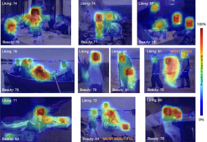
The video below shows animated eye movements while 20 subjects have viewed the paintings for 5 seconds each.
Eyes of Terror
Video showing eye movements from 28 viewers watching a scene from the horror movie “The Conjuring 2”. Red disks show gaze position for each viewer, and the heatmap shows the mean focus of gaze, that is, the region where most viewers are currently looking at
The Theory of Fear
In 2020 we teamed ups with Samsung Electronics Nordic to create a chilling horror experience for fans of thrills. When science meets immersive sound and picture, true horror can be created!
The Godfather synchronizes brains across individuals
Video highlighting main results for Nummenmaa et al (2012 PNAS); shows how brain activity becomes synchronized across inividuals exposed to unpleasant emotional content. The video was created by Emotion Lab ex-post-doc Enrico Glerean. Movie (The Godfather by Francis Ford Coppola) © Paramount pictures.
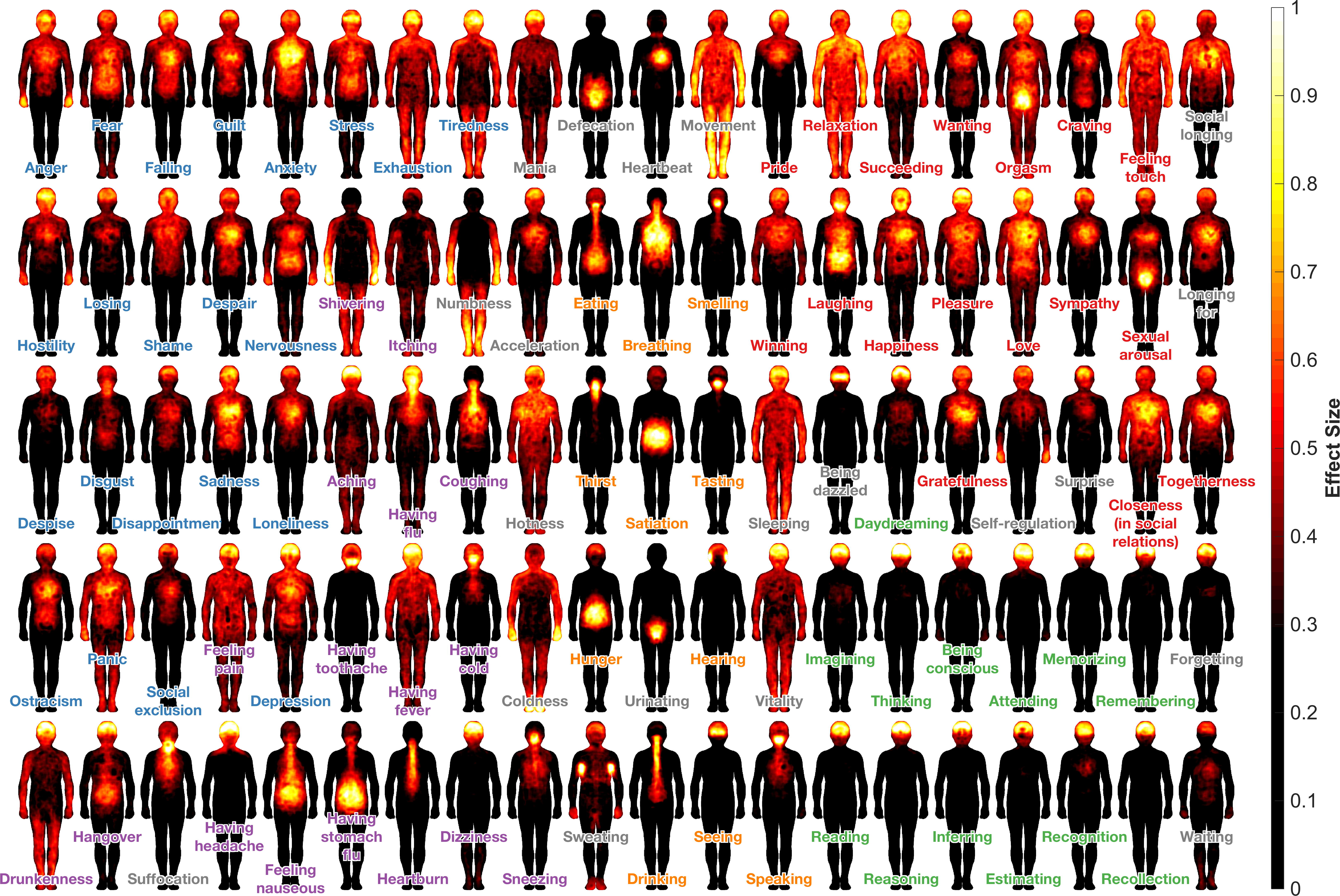
Maps of Subjective Feelings
This study mapped the presentation of 100 different feelings in the body. The figure below shows the mean sensation map for each feeling. A high-resolution version is available for download here. If you wish to translate the figure to another language, there’s a version without the labels available too; click here to download. Lay summary of the study (in English and in Finnish) can be accessed from the links below.
Kokemusmaailman kartta
Cartography of consciousness
The vide below shows animated version of each feeling. The video is CC-licensed and is also available in Finnish, Japanese, Spanish, and Italian. The video was created by Emotion Lab ex-post-doc Dr. Enrico Glerean.
Animated bodily maps of emotions
Main figure from Nummenmaa et al 2014 PNAS; shows bodily regions where different emotions are felt in adults. If you want to use the animations in your presentations, you can also load the individual body animations from this link.
Seek and Destroy
Both children, adolescents and adults enjoy playing video games although they do not have immediate survival function. This video shows sample gameplay from our study where we demonstrated consistent activity in the brain’s reward circuit while winning in a competitive first-person video game playing.
Animated Human Erogenous Zones
This video (Nummenmaa et al 2016 Arch Sex Behav) reveals topography of human erogenous zones whose touching feels arousing while masturbating or having sex with a partner. The video starts by showing all the erogenous zones, and proceeds towards showing only the most erogenous areas. Yellow regions have the highest and red regions the lowest capacity for triggering arousal. Regions without overlaid colouring lack erogenous capacity completely. A fun fact: This video was rated as too raunchy per YouTube so we cannot host it there. Luckily our own servers can tolerate hardcore science
Images
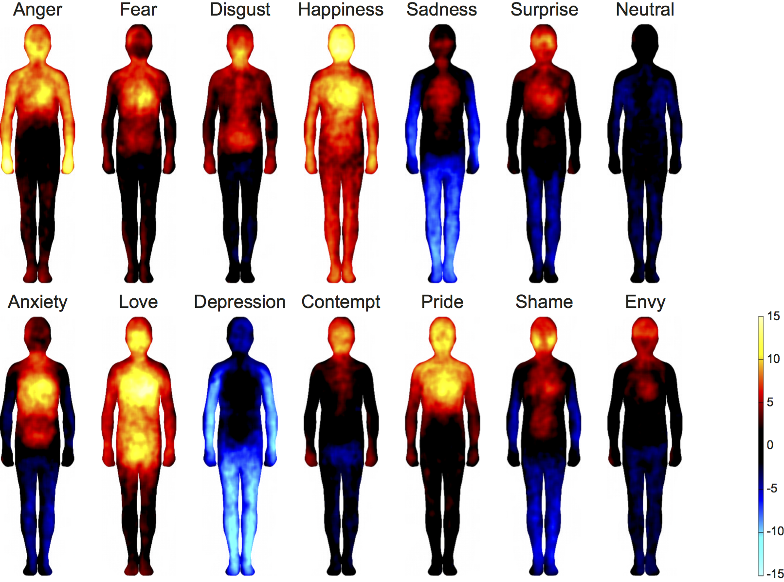
Bodily maps of emotions
Topographical organization of human emotions in the body from Nummenmaa et al (2014 PNAS). The data are based on self-reports of 400 adults. A version with Finnish labels can be downloaded here and an unlabelled version for translations is available here.
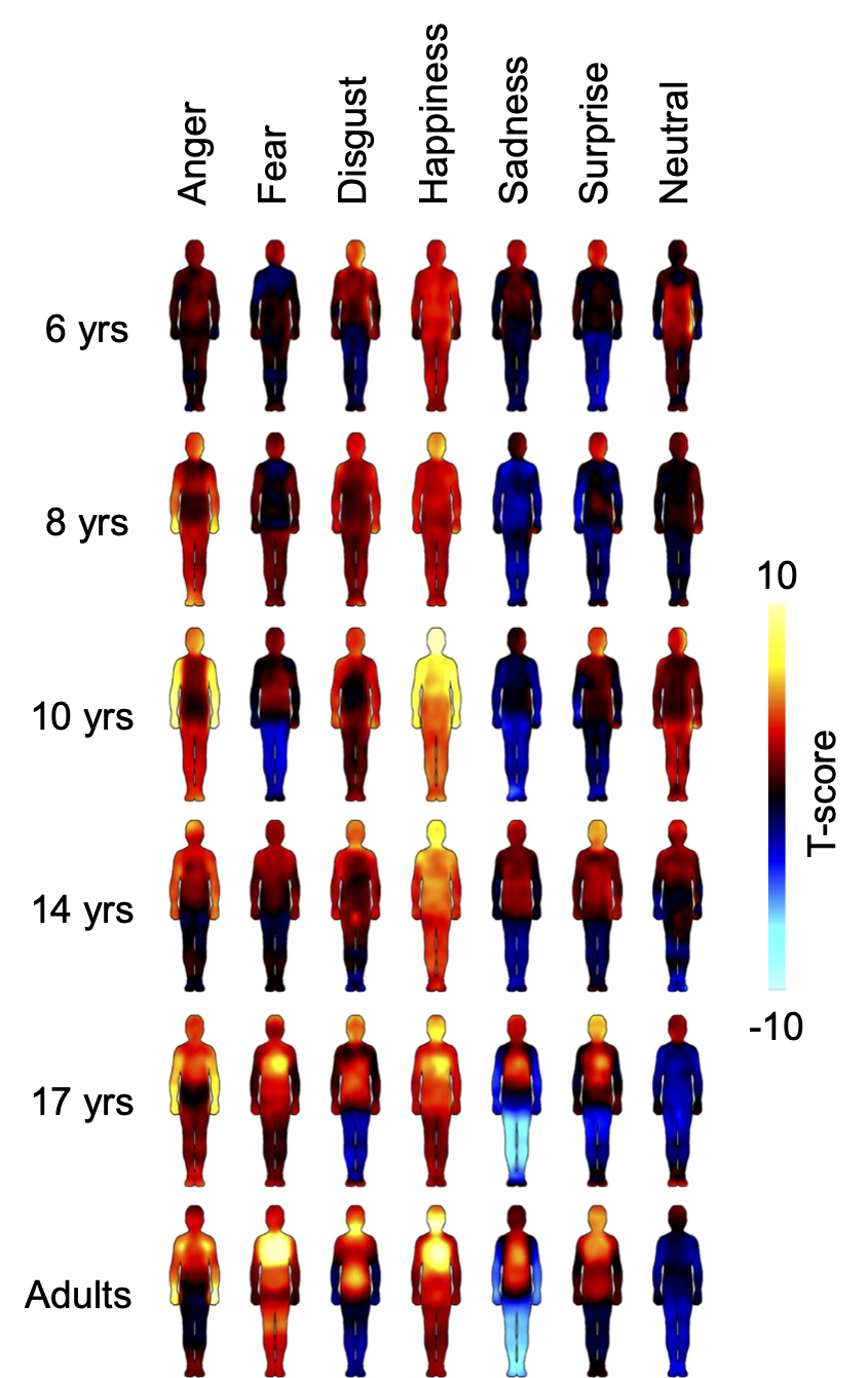
Bodily maps of emotions across child development
Main figure from Hietanen et al (2015 Developmental Science); shows bodily regions where different emotions are felt in children and adolescents.
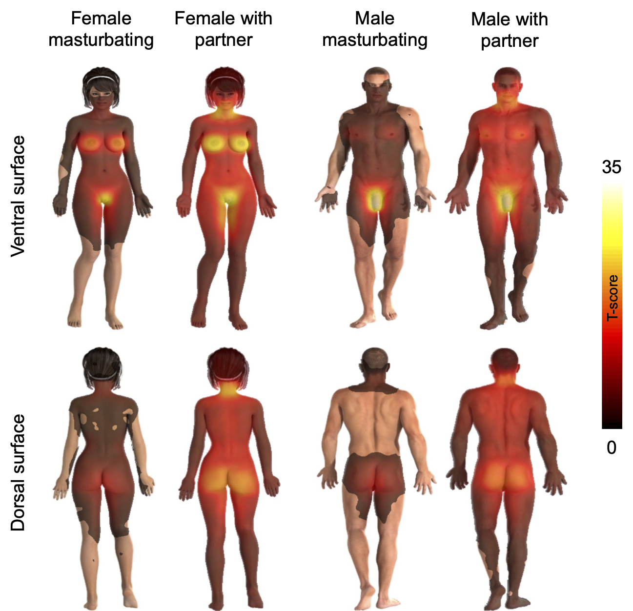
Human Erogenous Zones
Main figure from Nummenmaa et al 2016 (Arch Sex Behav) shows the topography of human erogenous zones while having sex with a partner and when masturbating. Yellow regions have the highest and red regions the lowest capacity for triggering arousal. Regions without overlaid colouring lack erogenous capacity completely. The data are based on reports from 700 adults.
Social touch zones
Main figure from Suvilehto et al 2015 PNAS; shows bodily regions where different memembers of one’s social networks may touch them.

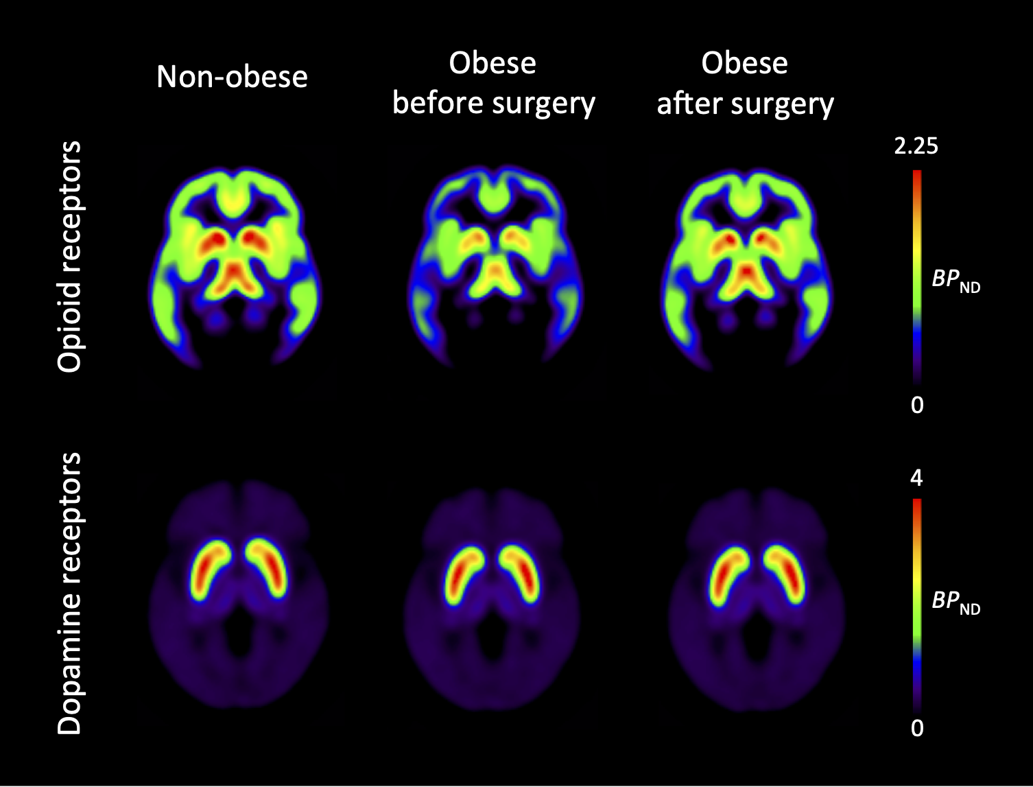
Obesity and brain
Main results from Karlsson et al (2015 Mol Psych) showing how obesity lowers endogenous opioid system tone, which, however, recovers following weight loss.