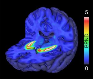
 Processing and statistical analysis of Brain-PET and (f)MRI data
October 4.-5. 2017 @Turku PET Centre
This is the webpage for the Turku PET Centre neuroimaging course organised in 2017. The course has ended, but all materials remain freely available. You can view PDF versions of the presentations by clicking on the lecture title.
Turku PET Centre neuroscientists are happy to annoucnce the first-ever PET-MRI neuroimaging course. This course will give a hands-on overview on the basic brain imaging analysis techniques applicable to PET as well as structural and functional MRI data. The course is aimed for PhD students, post-docs, and scientists working on medical, cognitive, and computational neuroimaging. Participants are assumed to have rudimentary knowledge of neuroimaging and working with PET and MRI data, but this is not strictly necessary. After the course, the participants should be able to process and analyze the data that has been retrieved from MRI scanner / PET camera independently. Analytic pipeline will focus on the PET Centre data analysis framework but can be adopted to any MRI / PET analysis platform.
The course is free but registration is mandatory. Registration is on first-come-first-served basis with priority to Turku PET Centre scientists and students. We can accommodate a maximum of 40 participants.
Registration for the course has been closed
FINAL PROGRAM
Wednesday October 4th: Brain imaging theory and preprocessing
09:00-10:00: Basics principles of human neuroimaging (Lauri Nummenmaa)
10:00-11:00: Quantifying brain structure and function with MRI (Virva Saunavaara)
11:00-12:00: Pharmacokinetic modelling of PET data (Jussi Hirvonen)
12:00-13:00: Lunch break
13:00-14:00: Preprocessing PET data with the PET Center pipeline (Tomi Karjalainen)
14:00-14:45: Preprocessing structural MRI: data with VBM and Freesurfer (Marco Bucci)
14:45-15:15: Coffee break
15:15-16:00: fMRI preprocessing (Enrico Glerean)
16:00-17:00: Preprocessing Diffusion Tensor Imaging data (Virva Saunavaara)
Thursday October 5th: Statistical analysis of brain imaging data
09:00-10:00: Statistical analysis of volume and surface-based data (Lauri Nummenmaa)
10:00-12:00: General Linear Model for random-effects fMRI (Linda Henriksson)
12:00-13:00: Lunch break
13:00-14:00: Connectivity analysis (Enrico Glerean)
14:00-15:00: Visualizing volume and surface based data (Tomi Karjalainen & Jouni Tuisku)
15:00-16:00: Wrap-up & General Q/A with coffee
Virtual lecture by Mr Malmsteen can be viewed here
LINKS TO SOFTWARE PAGES
Data analysis software and tools
SPM12: Multimodal (MRI / PET / EEG / MEG) data processing and analysis suite, runs on Matlab
FSL: Standalone MRI data processing and analysis suite
Freesurfer: Standalone tool for analysis and visualization of structural and functional neuroimaging. Most well known for the excellent cortical surface mesh generation algorithms
AFNI: Standalone MRI data processing and analysis suite
BrainVoyager: Standalone commercial MRI data processing and analysis suite
PMOD: Standalone commerical PET modelling and image processing tool
DTI tools
DTIprep: General-purpose too for DTI analysis
FSL: has also good tensor fitting packages as well as tools for tract-based analysis
ExploreDTI: a graphical toolbox for tractography
Volumometric image viewer tools
MRIcron: Image viewer with basic analytic, image manipulation and ROI drawing capabilities
Mango: Advanced volumometric viewer with image editing and rendering capabilities (http://ric.uthscsa.edu/mango/)
Carimas: Medical image analysis platform developed at Turku PET Centre. Capable of visualization, segmentation and modelling of multiple types of medical images
Freeview: Built-in viewer for Fressurfer. Excellent for viewing 2D and 3D images in tandem
FSLview: Built-in image viewer for FSL. Good capailities for volume viewing
3D rendering tools
Freeview: Built-in viewer for Fressurfer. Excellent for viewing 2D and 3D images in tandem
3dSlicer: Open-source software package for visualization and medical image computing
Caret: Multi-purpose surface visualization and analysis tool
MRIcroGL: Graphially more advanced and faster version of MRIcron with better 3D habdling capabilities
FSLview can also do some 3D image rendering